
A professional overview of how an optical system, a camera and a touchscreen turn a glass slide into an image that a whole group can study together.
Why a Screen Changes the Way Slides Are Studied
A conventional microscope shows a specimen to one person at a time. That slows case review, makes teaching harder and leaves little record of what was seen. A digital laboratory microscope adds a camera and a display, so the same view can be examined by several people, saved and attached to a report.
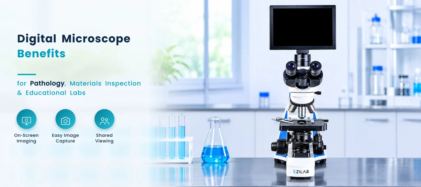
The Digital Microscope EZL-DM101 from Ezilab is a digital lab microscope with a free trinocular head, infinity plan objectives, a built-in LED lamp, a five megapixel camera and a touchscreen. This article explains how that kind of instrument works, where it is used and which selection mistakes are worth avoiding.
From Specimen to Screen: How the Image Forms
Light from the lamp passes through a condenser and the specimen, and the objective lens builds a magnified image. In an infinite optical system, the objective sends parallel light toward the head, which allows accessories such as filters and contrast kits to be added without degrading the image.
In a digital microscope laboratory setup, the head has a third port. One path feeds the eyepieces for direct viewing, and the other feeds a camera. The camera sensor converts the image into digital data, which the screen displays and which can be saved or shared.
Magnification is the product of the objective and the eyepiece. Here, objectives of four, ten, forty and one hundred times work with ten times eyepieces, and the page does not state a single total figure.
Reading a Digital Microscope Diagram, Part by Part
A simple digital microscope diagram shows why the image path is easy to follow. Light passes through the specimen, the objective forms the image, and the head splits it between the eyepieces and the camera.
Diagram: light and image path, parts lettered A to F
- AIllumination — A built-in LED lamp and an Abbe condenser light the specimen.
- BStage — A double-layer mechanical stage moves the slide smoothly.
- CObjectives — Four infinity plan objectives sit on a quadruple nosepiece.
- DEyepieces — Wide field plan eyepieces give direct viewing through the trinocular head.
- ECamera — A five megapixel camera captures the image.
- FScreen and output — A touchscreen shows the image, and a card slot, USB, HDMI and Wi-Fi save or share it.
Examining a Slide Step by Step
In daily use, a digital microscope for laboratory work follows a short routine.
Sharp Imaging, Easy Capture and Shared Viewing
Sharp On-Screen Imaging
Infinity plan objectives, wide field plan eyepieces and a five megapixel camera support a clear view.
Easy Image Capture
Images save to a card or export through USB, HDMI or Wi-Fi for use in reports.
Comfortable Shared Viewing
A nine point seven inch touchscreen lets groups review a slide together instead of taking turns at an eyepiece.
Many Contrast Methods
One stand covers brightfield, phase, dark field, fluorescence and live-cell viewing with the supplied kits.
Precise Focus
Coaxial coarse and fine focus has a fine division of two thousandths of a millimeter.
Flexible Sharing
Built-in Wi-Fi and HDMI support case review sessions across a room.
EZL-DM101 Technical Details
| Parameter | Value |
|---|---|
| Optical system | Infinite, with infinity plan objectives |
| Objectives | Four, ten, forty and one hundred times, on a quadruple nosepiece |
| Eyepieces | Wide field plan, ten times |
| Head | Free trinocular, inclined thirty degrees |
| Interpupillary distance | Forty-eight to seventy-five millimeters |
| Condenser | Abbe, numerical aperture one point two five |
| Stage | Double-layer mechanical, one hundred forty by one hundred forty millimeters |
| Camera | Five megapixels |
| Display | Nine point seven inch touchscreen running Android |
| Software | S-EYE |
| Output | Card slot, USB, HDMI and Wi-Fi |
| Illumination | Built-in LED |
| Size | Three hundred by three hundred by six hundred millimeters |
| Net weight | Ten kilograms |
The full list is available on the Digital Microscope EZL-DM101 product page.
Where It Fits: Pathology, Research and Teaching
Clinical Laboratories
Blood smears, urine sediments and stained tissue sections.
Research Laboratories
Cells and microorganisms.
Pathology Teams
Documenting findings for records and reports.
Schools, Colleges and Training Centers
Classroom demonstrations that a whole group can see.
Live Specimen Work
Phase contrast, dark field and a heating stage for living samples.
Case Review Sessions
Images shared over Wi-Fi or HDMI.
These digital microscope uses share one benefit: all present see the same field at the same time. A science lab microscope for teaching and a clinical unit differ mainly in the specimens, and readers who want to see the wider range can browse the digital microscope category page.
Contrast Methods Compared
| Method | Suited To | Supplied With This Model |
|---|---|---|
| Brightfield | Stained slides such as blood smears and tissue sections | Built-in LED and Abbe condenser |
| Phase contrast | Living, unstained cells | Phase contrast kit |
| Dark field | Small, low-contrast objects against a dark background | Wet and dry dark field condensers |
| Fluorescence | Specimens labeled with fluorescent dyes | Blue-band fluorescence kit |
This table gives general background on each method; the right choice depends on the specimen and the question being asked.
Common Selection Mistakes
Judging by camera megapixels alone. Optics decide detail, so look at objectives, eyepieces and the optical system as well.
Ignoring room lighting. Fluorescence and dark field images fade in a bright room, so plan a dimmable area.
Overlooking output needs. Decide whether images go to a card, a cable or Wi-Fi before choosing a model.
Forgetting the head type. A trinocular head allows both direct viewing and camera capture at once.
Skipping the contrast kits. Phase, dark field and fluorescence extend what the same stand can show.
Underestimating bench space. The unit measures six hundred millimeters tall and weighs ten kilograms net.
Habits That Keep Images Clear
- Clean lenses gently with proper lens tissue.
- Begin each session with the lowest objective.
- Adjust the condenser for each objective.
- Name saved images with a clear sample reference.
- Cover the instrument after use to keep dust out.
Exploring the Wider Family of Digital Microscopes
This model belongs to a category of microscopes for detailed observation and specimen examination in laboratory and research settings. Common features across it include a wide-field eyepiece to reduce eye strain, a dual-layer mechanical stage, an antifungal objective lens for humid environments and coaxial focus adjustment.
When reviewing a digital microscope or comparing options, readers usually look at objectives, eyepieces, head type, camera, display and output. The full range is easy to browse on the Ezilab digital microscope range, and wider laboratory categories sit on the Ezilab.
Continue to the Product Details
See the full specification list, accessories and applications for this model.
Explore Digital Microscope EZL-DM101Amelia Brooks
Blog Content Writer at Ezilab Scientific
Amelia Brooks is a blog content writer at Ezilab Scientific, specializing in laboratory equipment, scientific instrumentation, and technical content development. She focuses on translating complex product specifications and scientific concepts into clear, informative, and engaging articles. Her writing highlights equipment features, practical applications, and industry developments to help readers understand laboratory technologies and make informed decisions based on their technical requirements.