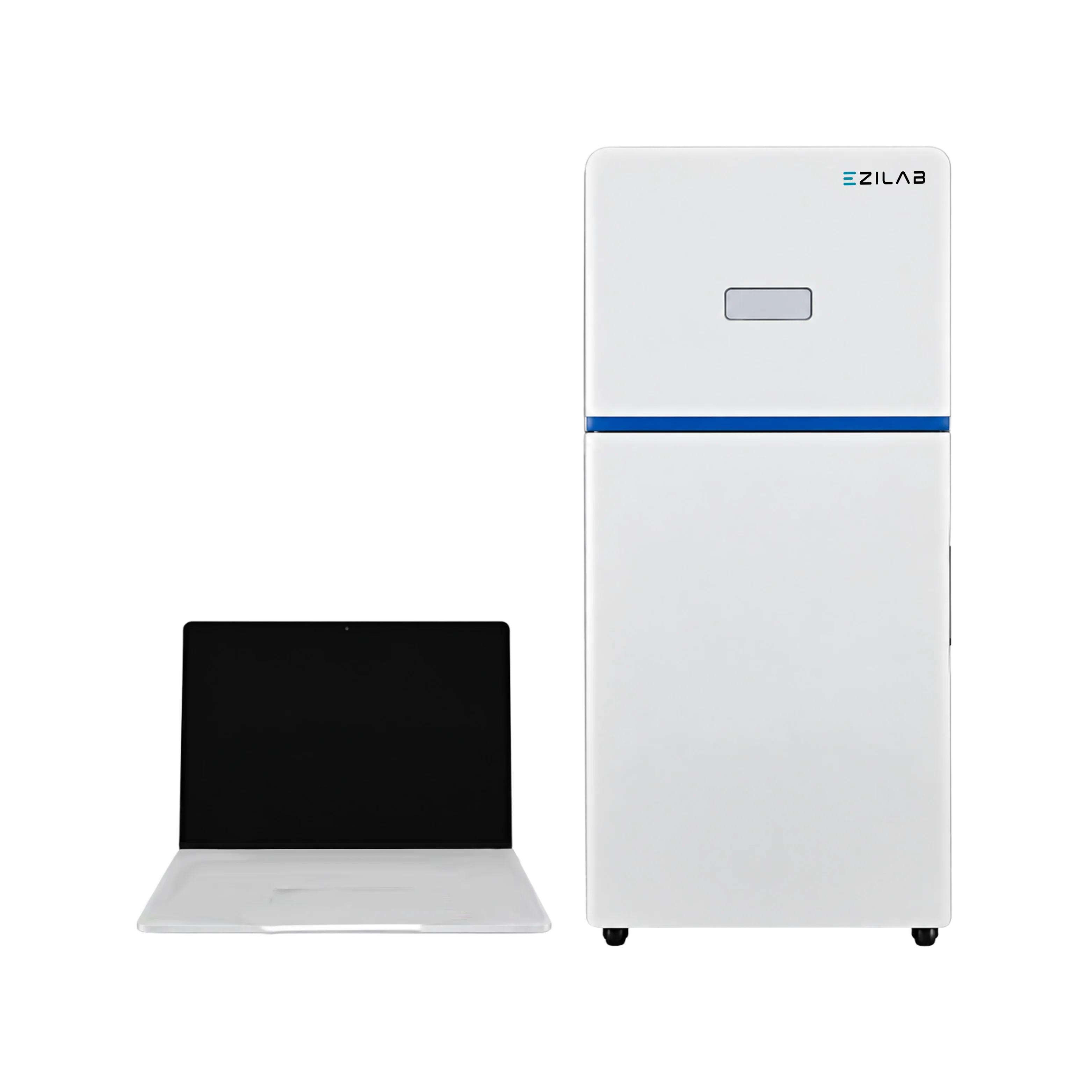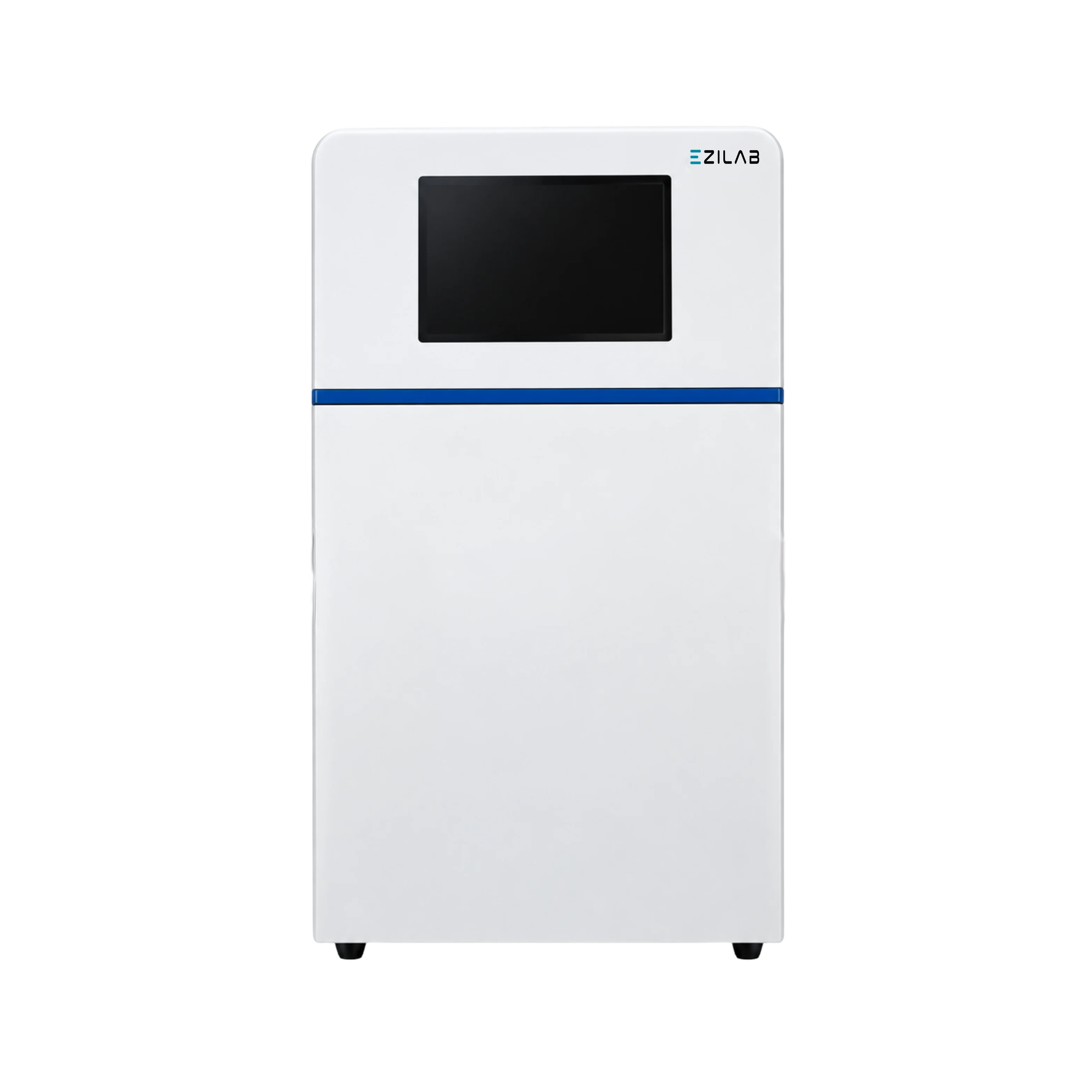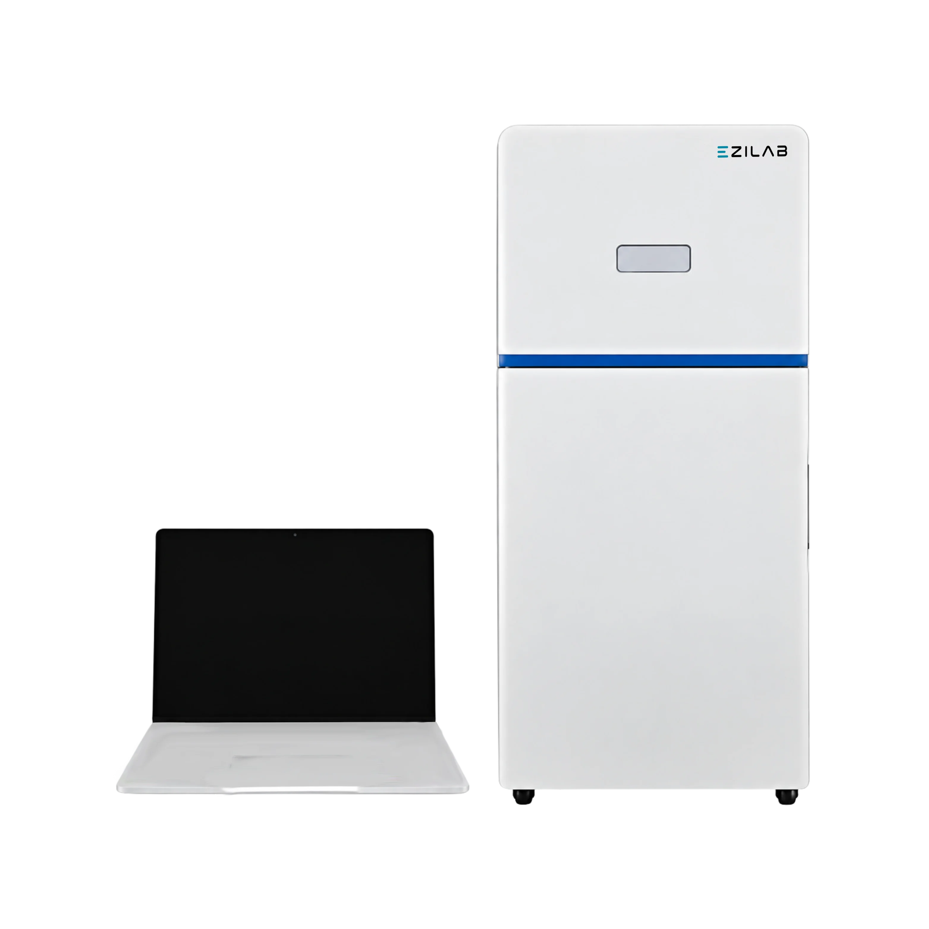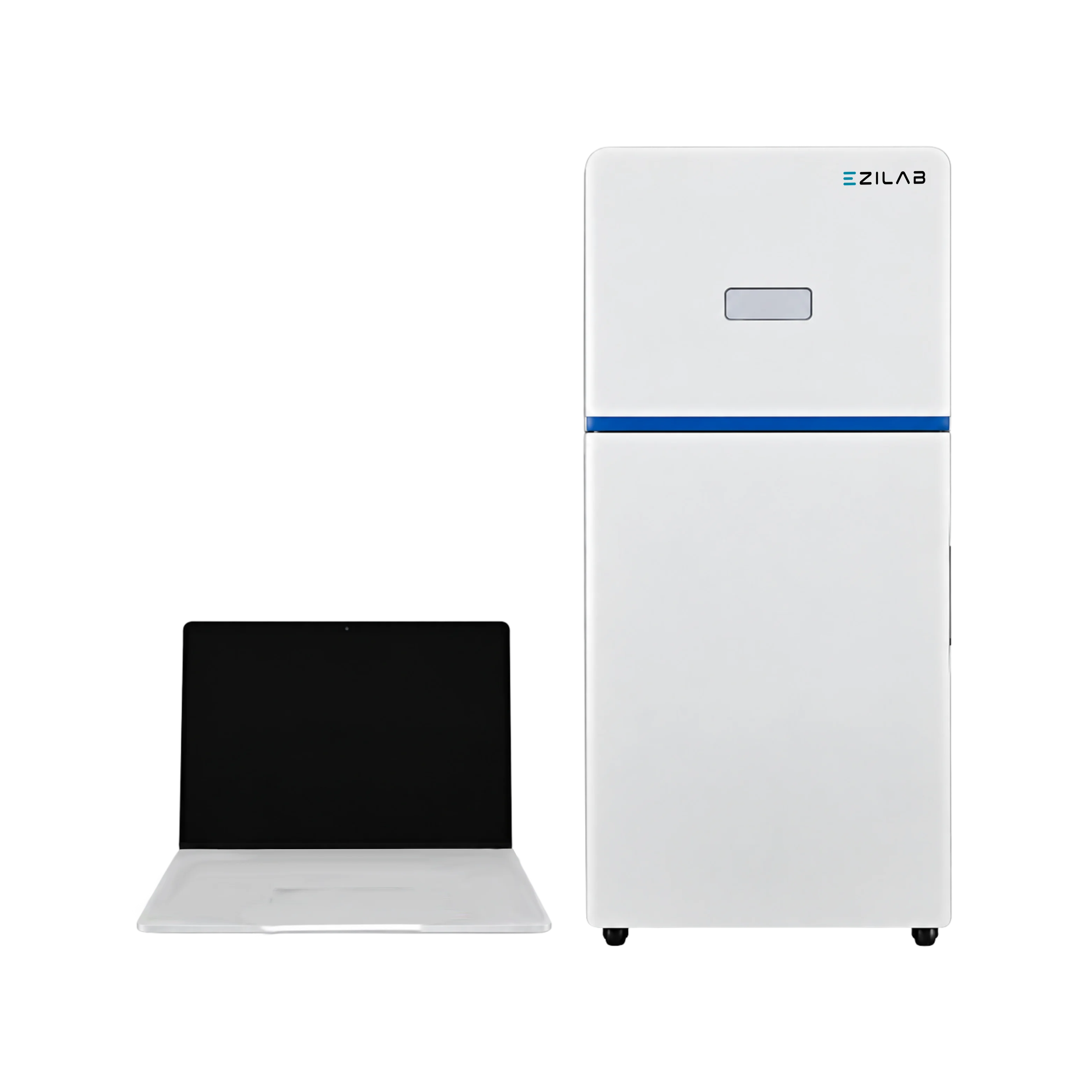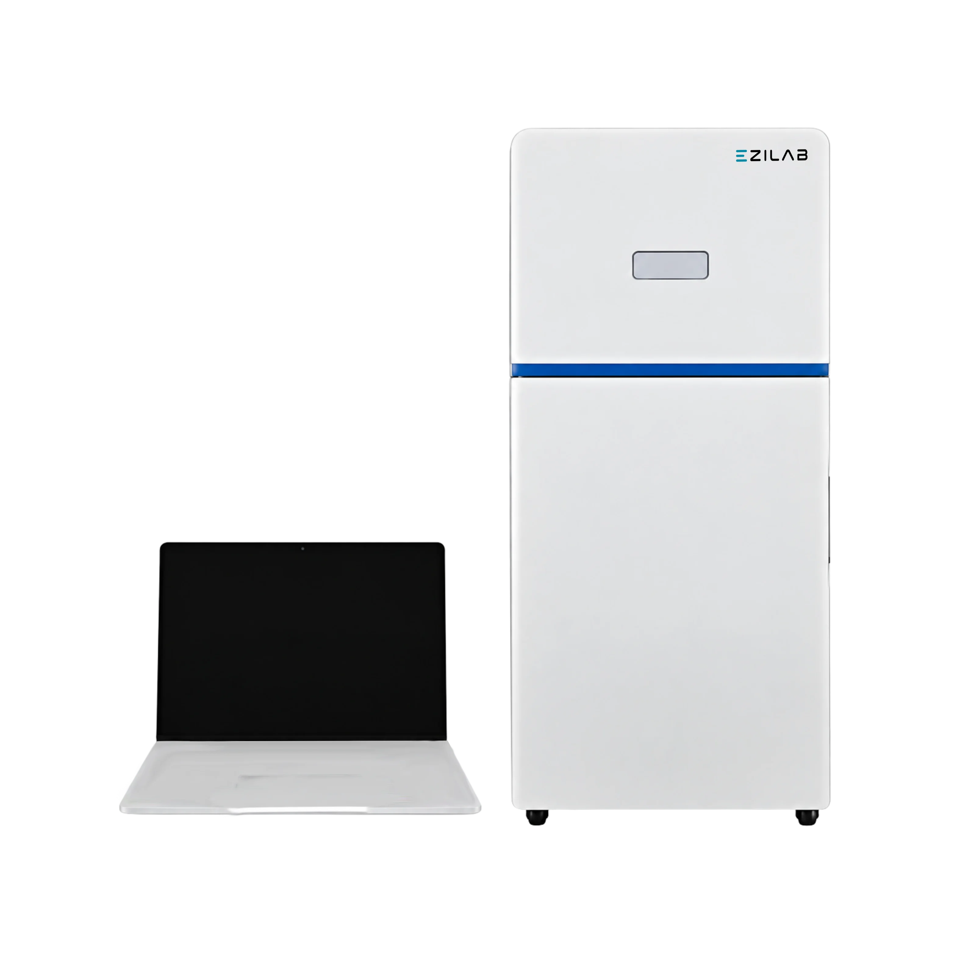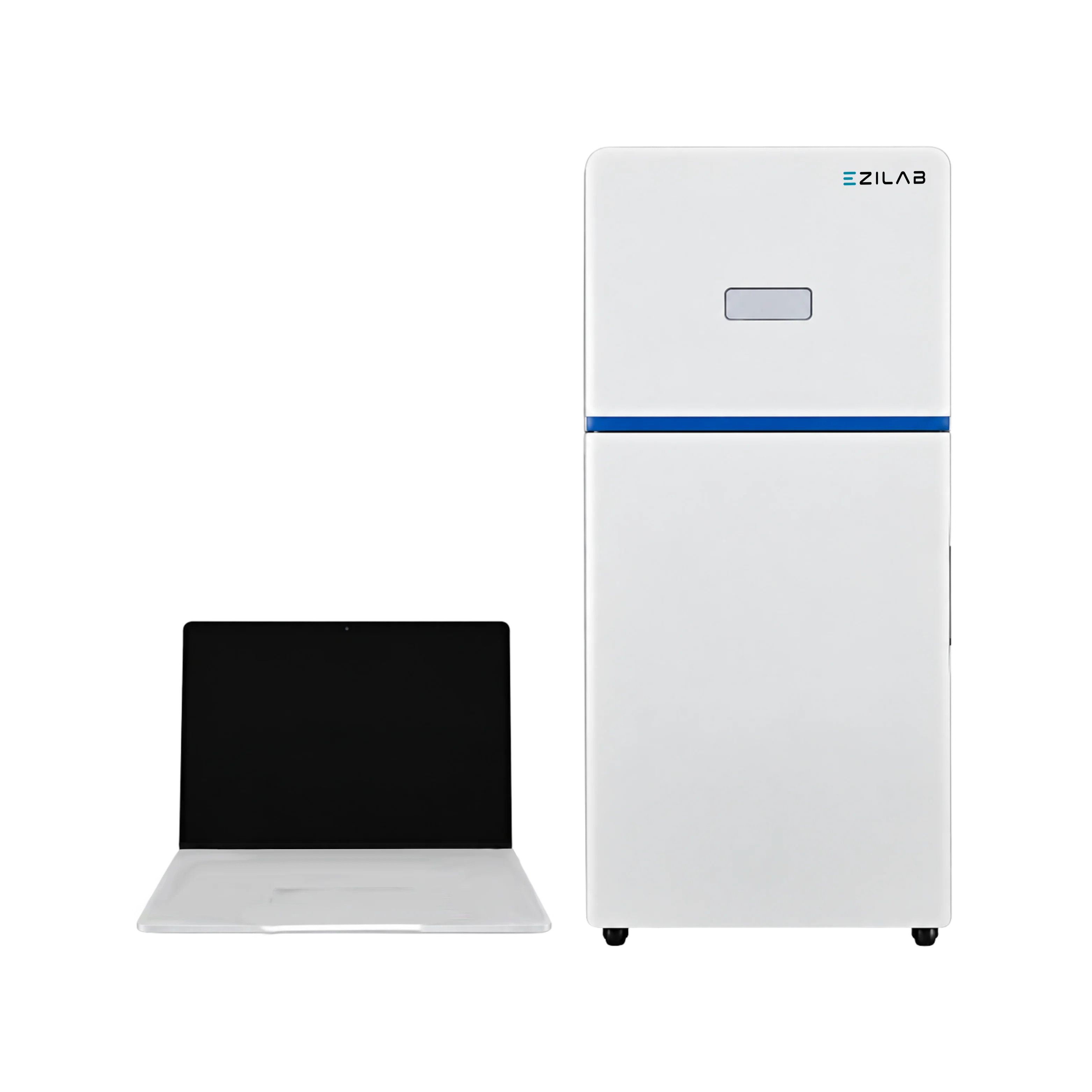
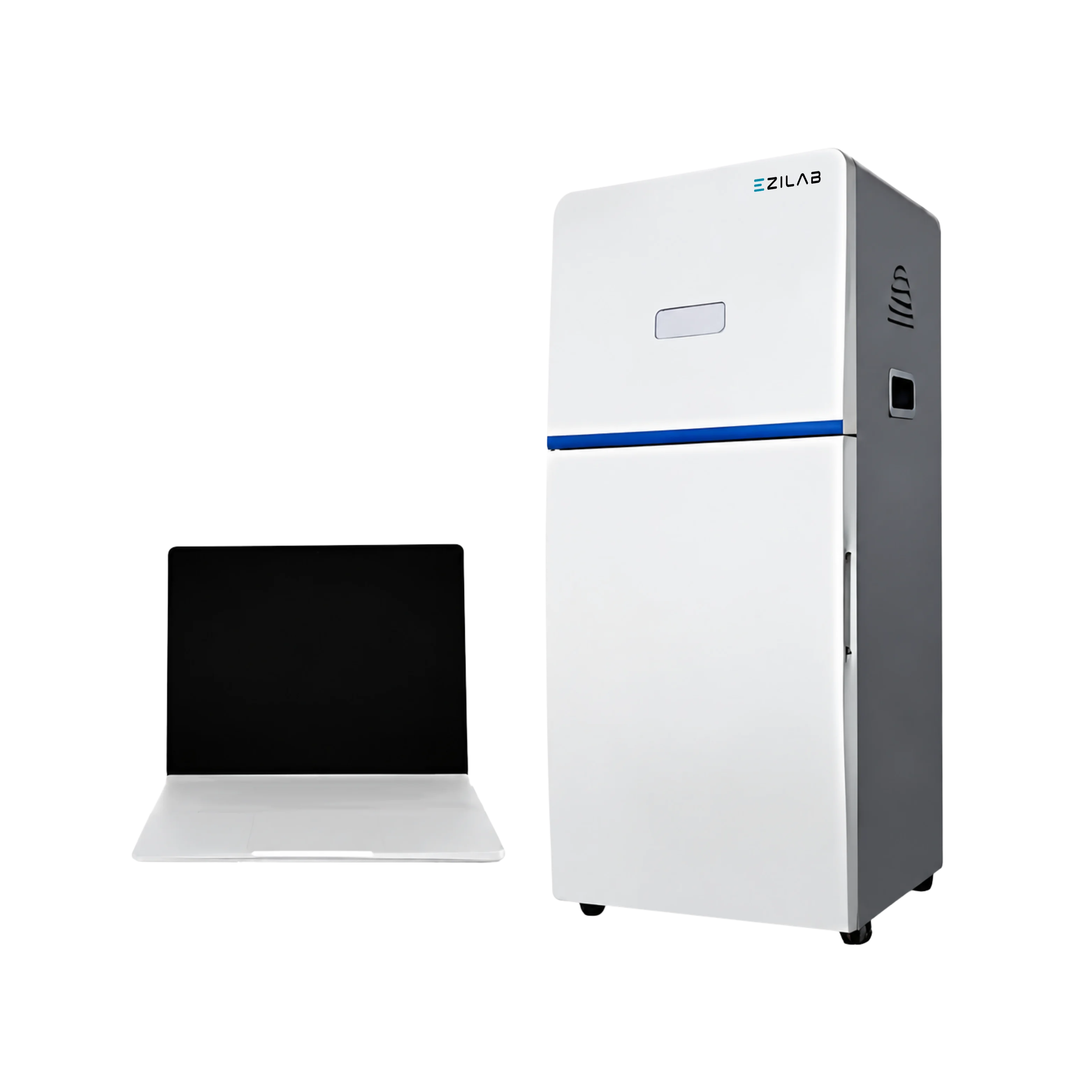
EziLab’s Gel Documentation Imaging System EZL-GD131 pairs a 5.0 megapixel scientific digital camera with 16-bit, 65536-grayscale depth and a 70.1dB signal-to-noise ratio, giving this gel doc system wider dynamic range than 8-bit sensors for faint-band detection. A UV smart trans-illuminator plus blue and white light options, automatic exposure, and pixel binning technology adapt the system to stained gels, membranes, and plates without manual guesswork, while built-in cropping, rotation, and color correction speed image cleanup before export. Optical density calculation supports quantitative band analysis from captured images. As a gel imaging system built for daily molecular biology work, this unit documents agarose and polyacrylamide gels, protein stains, and TLC plates on one camera platform. Molecular biology labs use it as a uv transilluminator alternative for routine gel inspection, while research groups running western blot imaging system workflows capture faint bands in one exposure. Microbiology labs apply the same platform as a colony counter for Petri dish counts, and ELISA plate imaging rounds out daily use. Academic core facilities standardize on shared imaging stations like this one for several busy research groups at once. See the Imaging System and Gel Doc Imaging System pages for further specifications and ordering details.
| Camera | Scientific Digital Camera |
|---|---|
| Resolution | 5.0 Megapixels, 2592×1944 |
| A/D | 16 bit ( 65536 Grayscale) |
| Pixel Density | 16 bit ( 65536 Grayscale) |
| Quantum Efficiency | ≥ 70% |
| Read Out Noise | 5.1e-RMS |
| Dark Current | e-/pixel/sec 25°C |
| SNR | 70.1db |
| Product Dimension (W×D×H) | 430×380×690 mm |
| Outer Pack Dimension (W×D×H) | 550×490×760 mm |
| Gross Weight | 31 kg |
Polymer by RIM molding
Better user experience
Switchable operation mode
UV smart trans illuminator
Blue and white light trans illuminator
Intelligent workflow
User interface allows total customization over imaging protocols
High precision automatic exposure
Advanced pixel binning technology
Automatic access of capture parameters
Image cropping, rotation and counter color process
Convenient image navigation and browsing
Calculation of optical density for the quantitative analysis
Optimization of the visual effects by wiping background mode
| • | Used in molecular biology laboratories for agarose and polyacrylamide gel inspection. |
| • | Applied in biochemistry laboratories for protein stain and membrane documentation. |
| • | Used in western blot imaging for capturing faint bands in a single exposure. |
| • | Applied in microbiology laboratories for Petri dish colony counting. |
| • | Used in analytical laboratories for TLC plate and ELISA plate imaging. |
| • | Applied in academic core facilities for shared imaging and optical density analysis. |