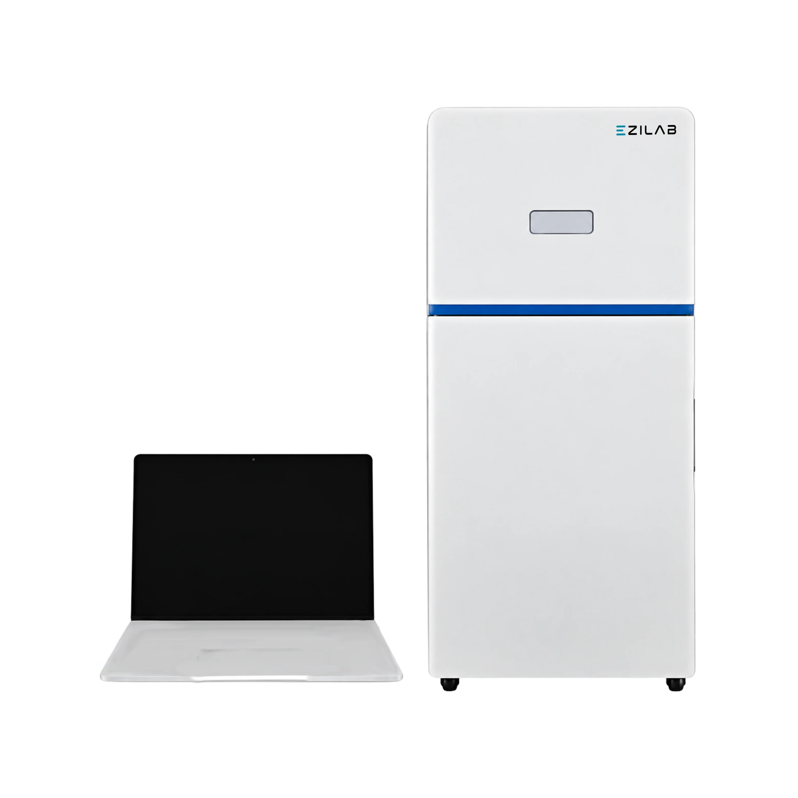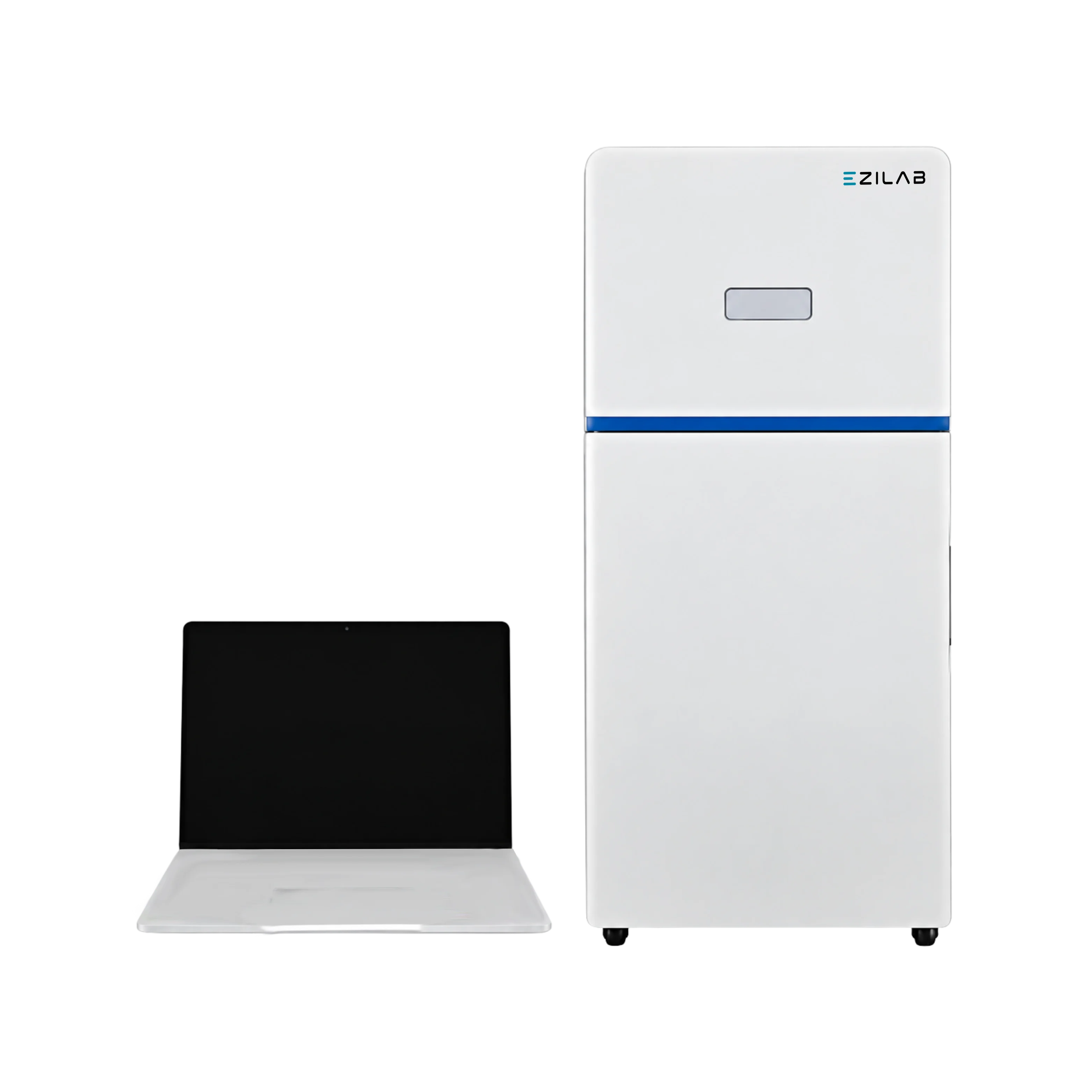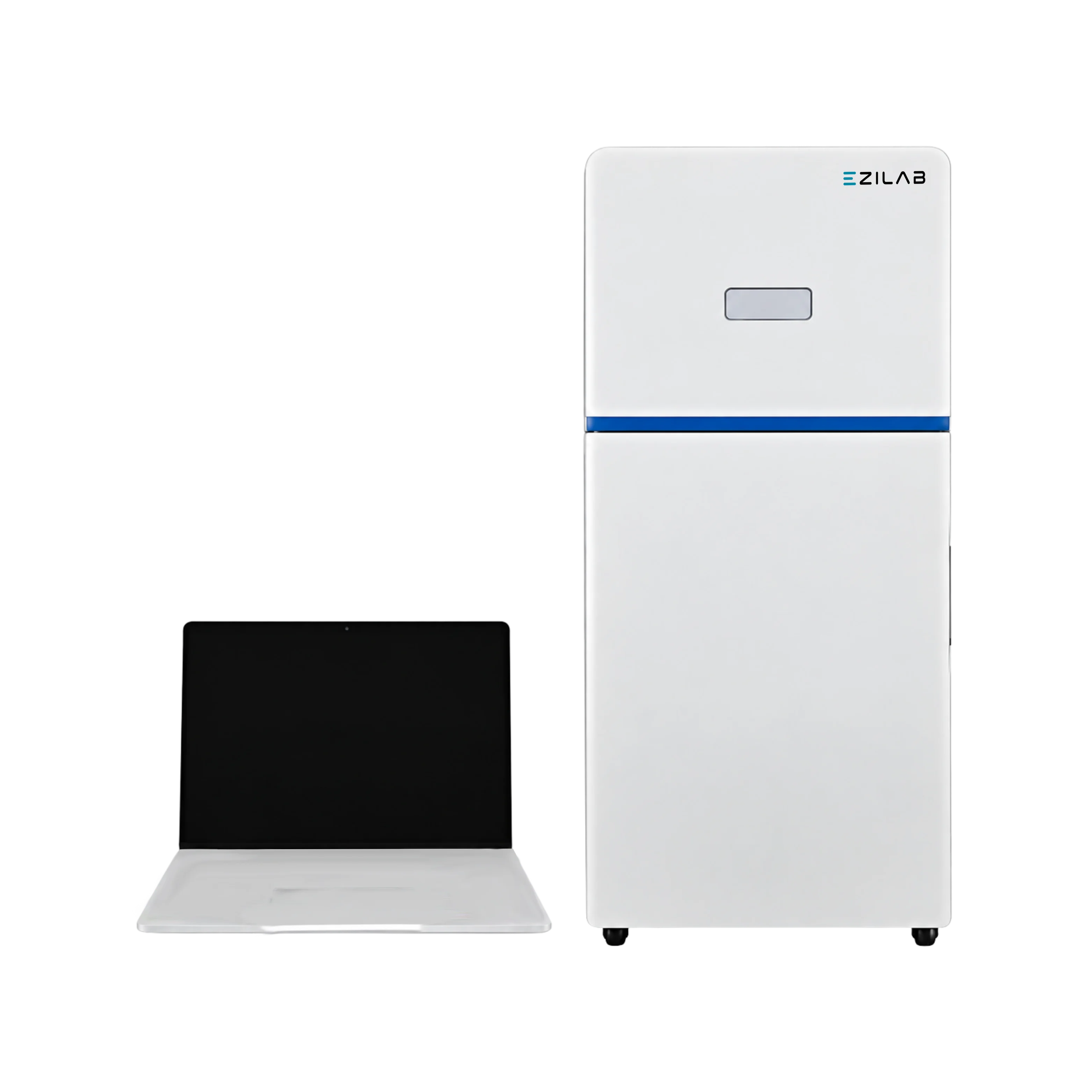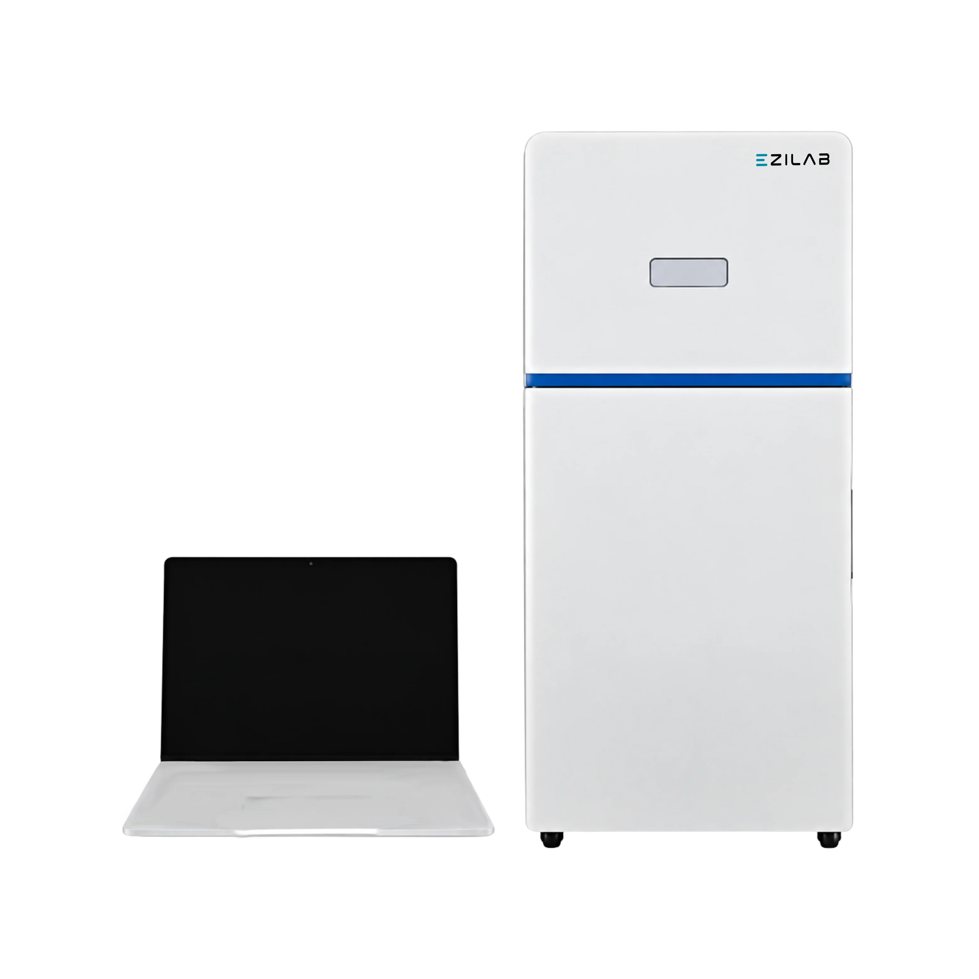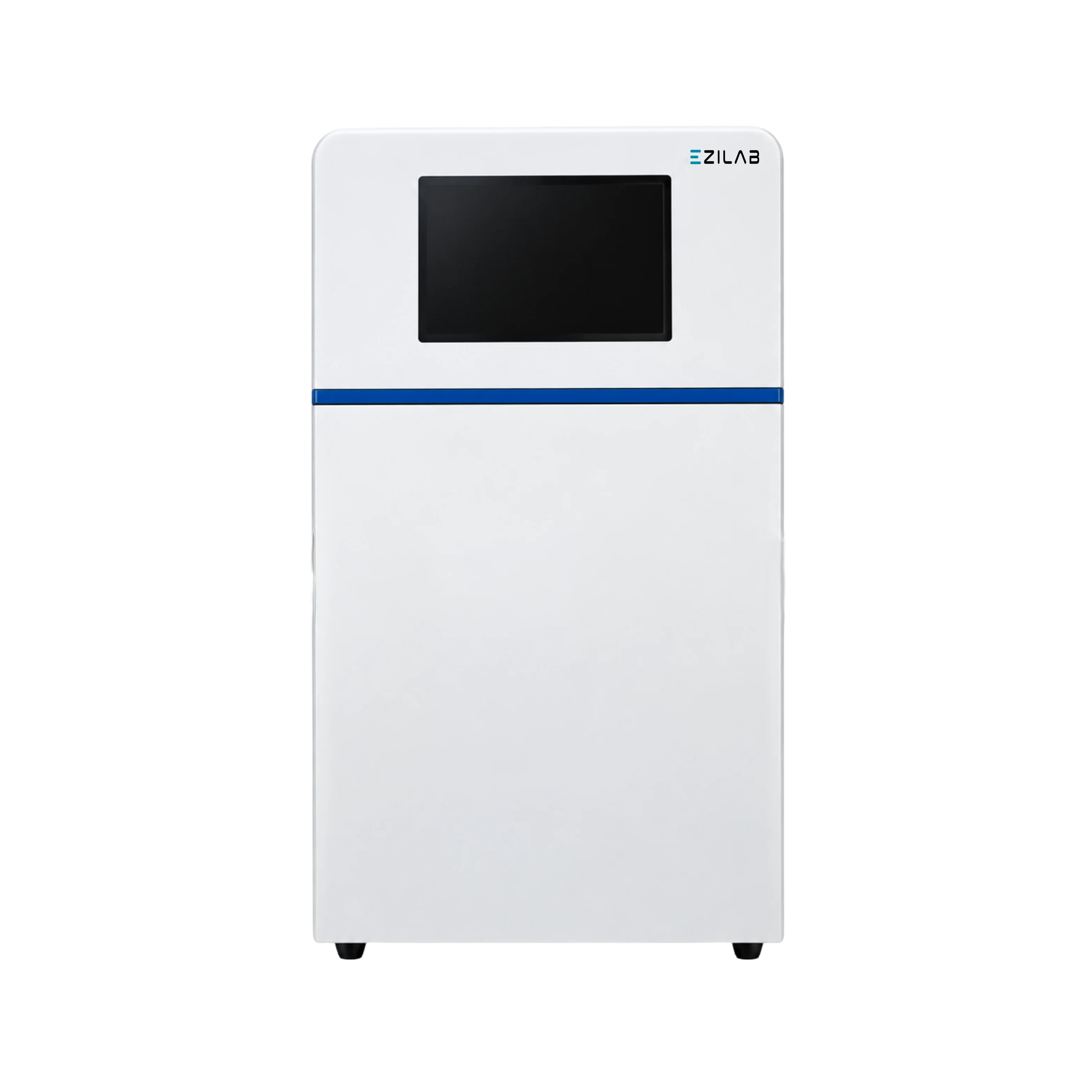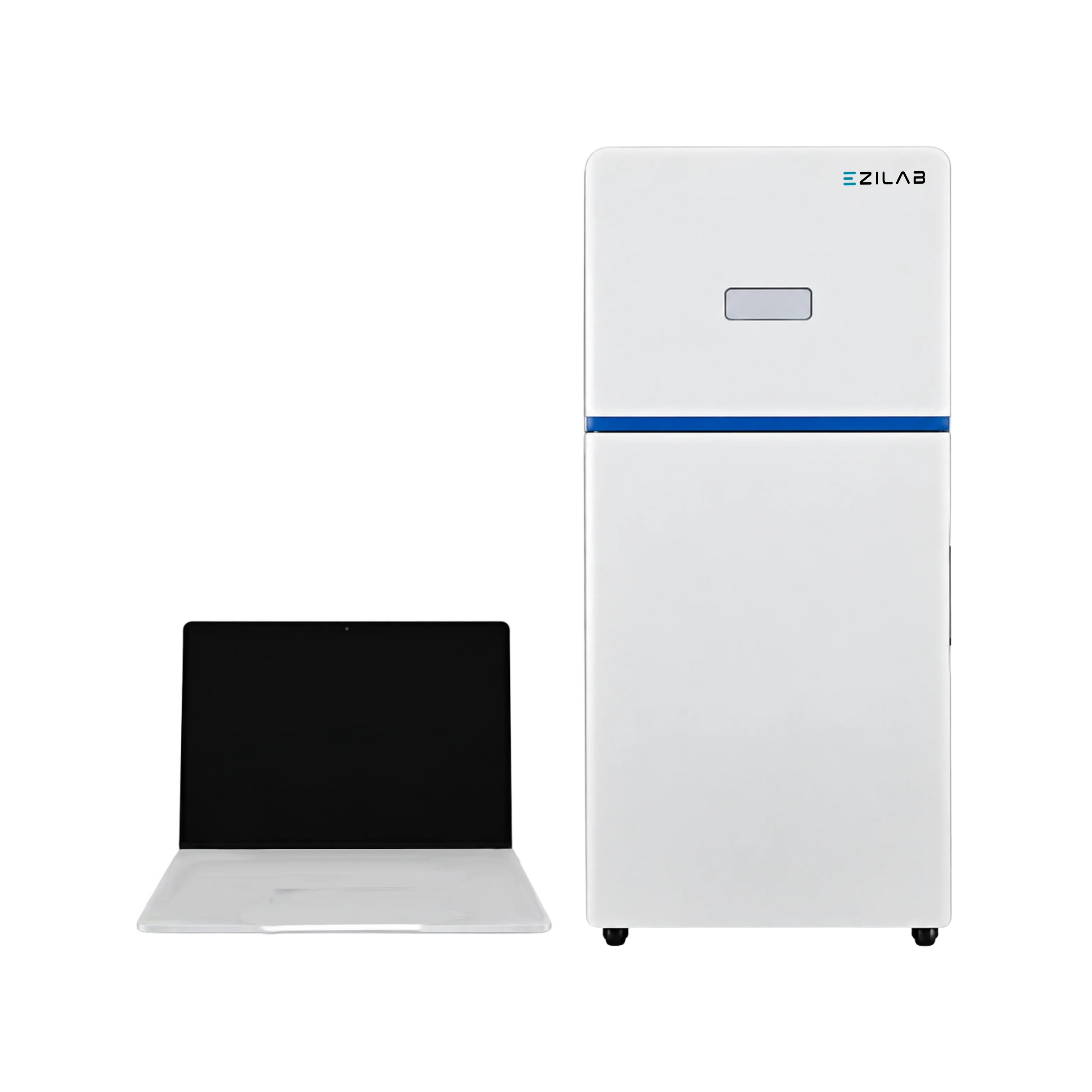
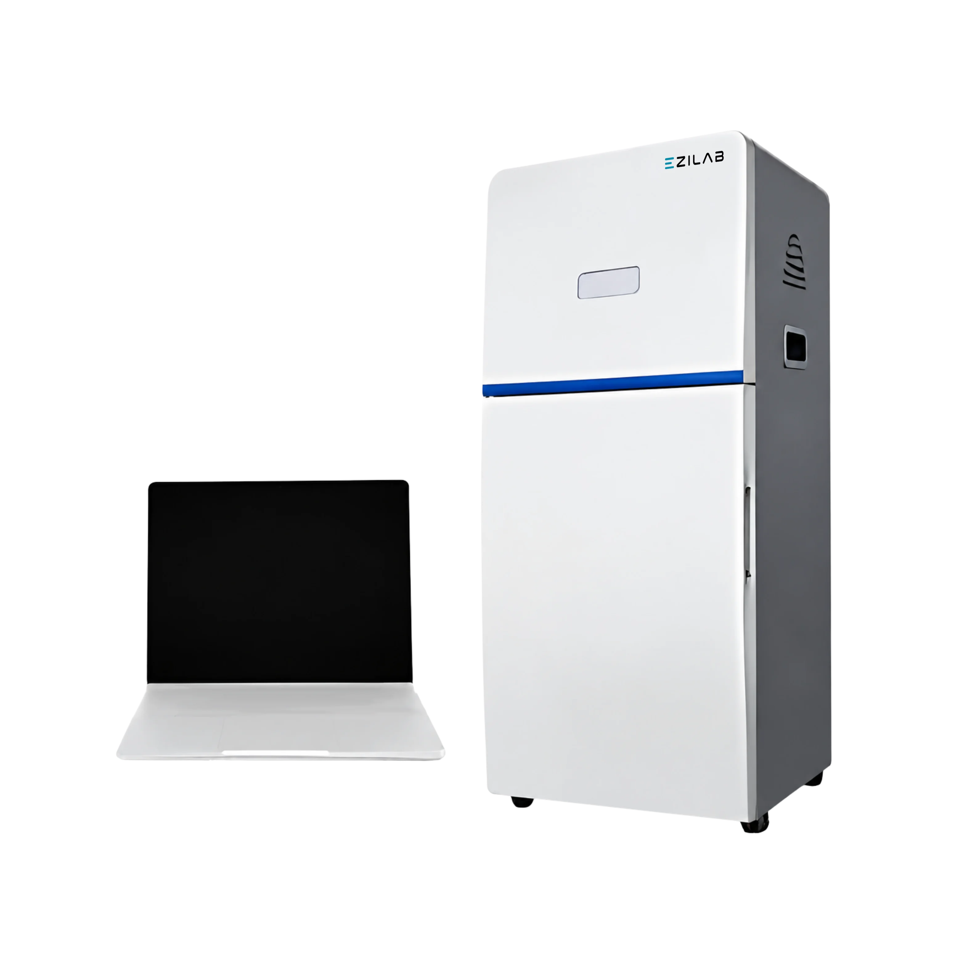
Ezilab's Chemiluminescence Imaging System EZL-CS133 serves molecular biology laboratories that need cooled CCD detection for capturing low-intensity chemiluminescent signals from Western blots, ELISA plates, and other luminescence-based assays. With a 6.05MP CCD sensor cooled to -25°C and touchscreen operation, this imaging system detects the faint light emissions chemiluminescent substrates produce during antibody-antigen or enzyme-based detection reactions, converting signals too weak for standard photography into quantifiable digital images. That -25°C CCD cooling is the critical spec here — cooling the sensor substantially reduces the electronic noise (dark current) that would otherwise obscure faint chemiluminescent signals, allowing detection of low-abundance proteins or weak reactions that an uncooled or lightly cooled camera would miss entirely against background noise. The 6.05MP resolution provides sufficient detail to distinguish closely spaced bands on a Western blot or resolve individual spots across a multi-well plate, while touchscreen operation simplifies exposure time adjustment and image capture settings compared to computer-only control interfaces. Molecular biology labs running protein detection work — Western blotting, ELISA-based chemiluminescent assays, and similar low-light imaging applications — get sensitive, quantifiable results from this cooled-sensor configuration. EziLab's Imaging System and Chemiluminescence Imaging System cover low-light molecular biology imaging needs across USA laboratory settings.
| Lens | Motorized F0.95 Ultrafast Lens |
|---|---|
| Touch Screen | 10.1 Inch Touch Screen |
| Camera | Scientific CCD Camera |
| Resolution | 6.05 Megapixels, 2750×2200 |
| Sensor Size | 12.49mm ×9.99mm |
| Pixel Density | 16 bit ( 65536 Grayscale) |
| Pixel Size | 4.54×4.54µm |
| Quantum Efficiency | ≥ 75% |
| Absolute Cooling Temperature | -25°C |
| Product Dimension (W×D×H) | 430×380×690 mm |
| Outer Pack Dimension (W×D×H) | 550×490×760 mm |
| Gross Weight | 31 kg |
Ultra slim trans illuminator
Front and door are designed by reaction injection molding
Better user experience
Switchable operation mode
Blue and White light trans illuminator
Intelligent workflow
User interface allows total customization over imaging protocols
High precision automatic exposure
Marker image capture and composition with objective brands
Convenient image navigation and browsing
Automatic recognition of gel bands
Optimization of the visual effects by wiping background mode
Advanced pixel binning technology
Calculation of optical density for the quantitative analysis
This system is widely used for Gel inspection, Protein inspection, Blotting Membrane, Color imaging and DNA/RNA analysis.