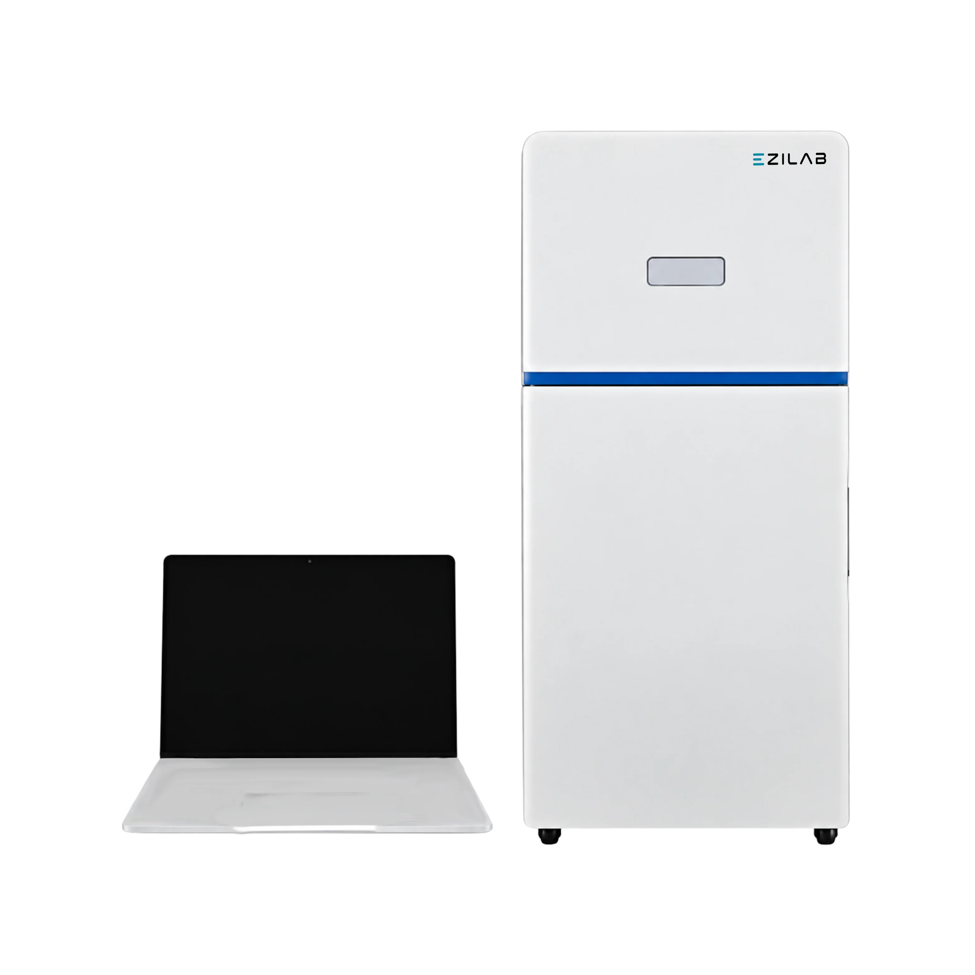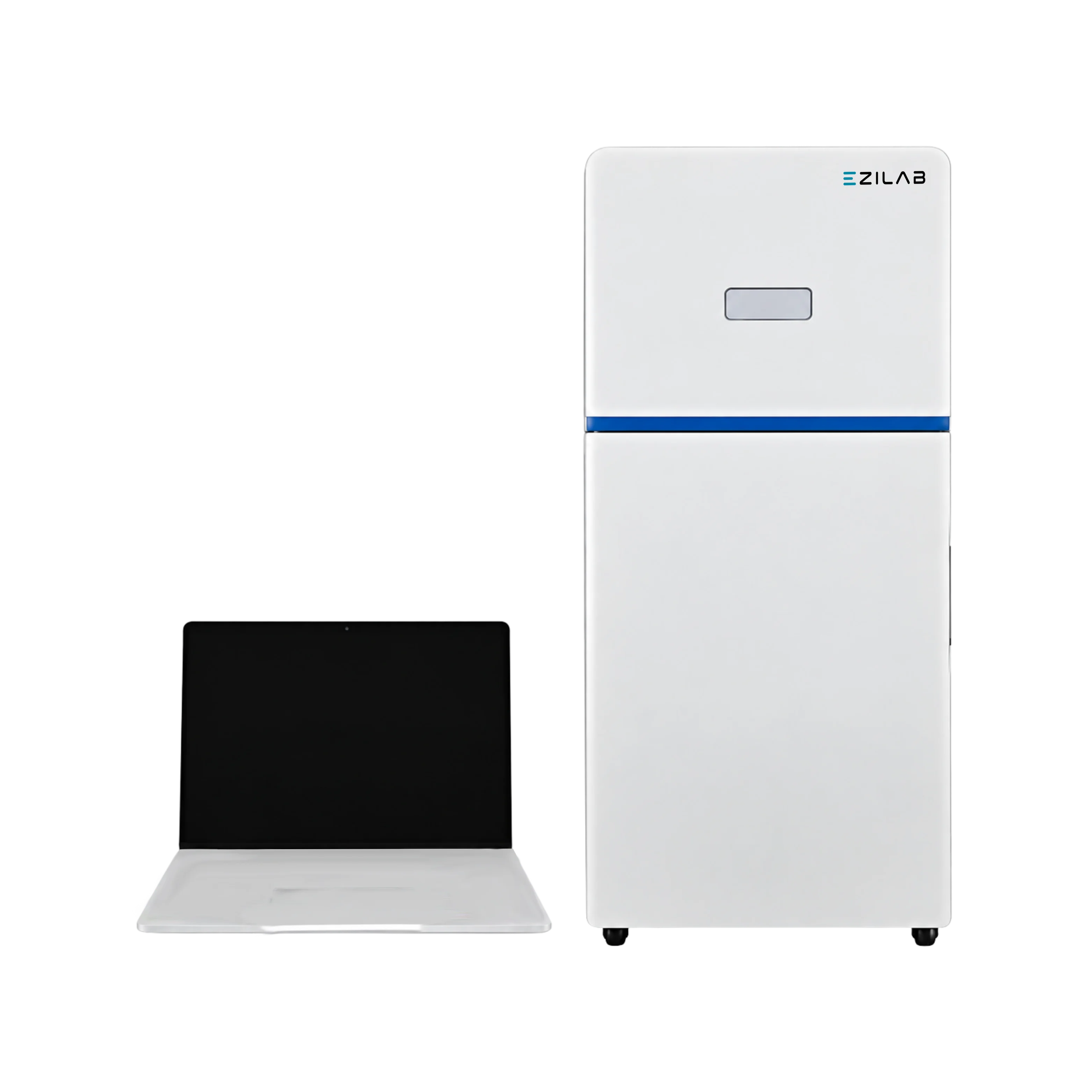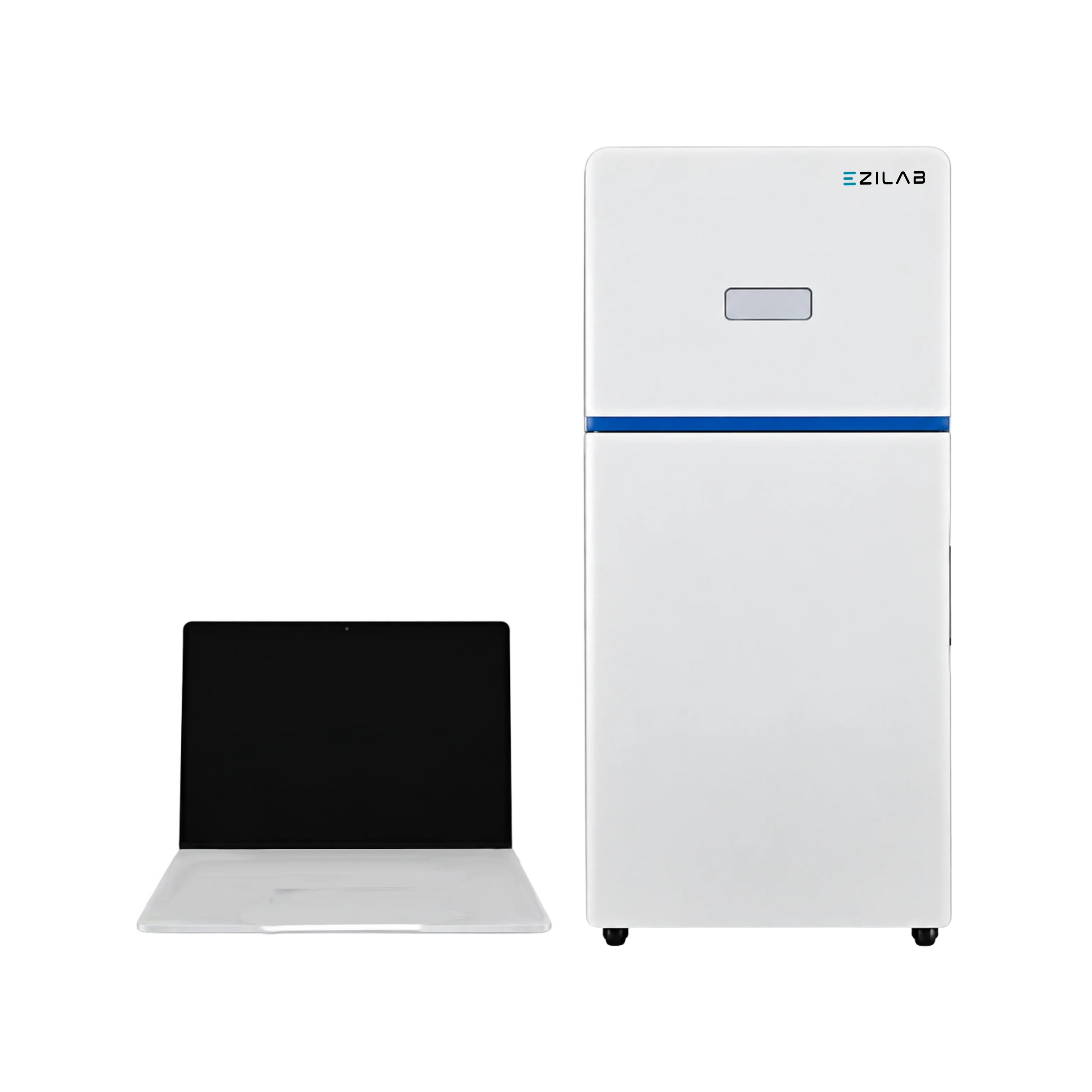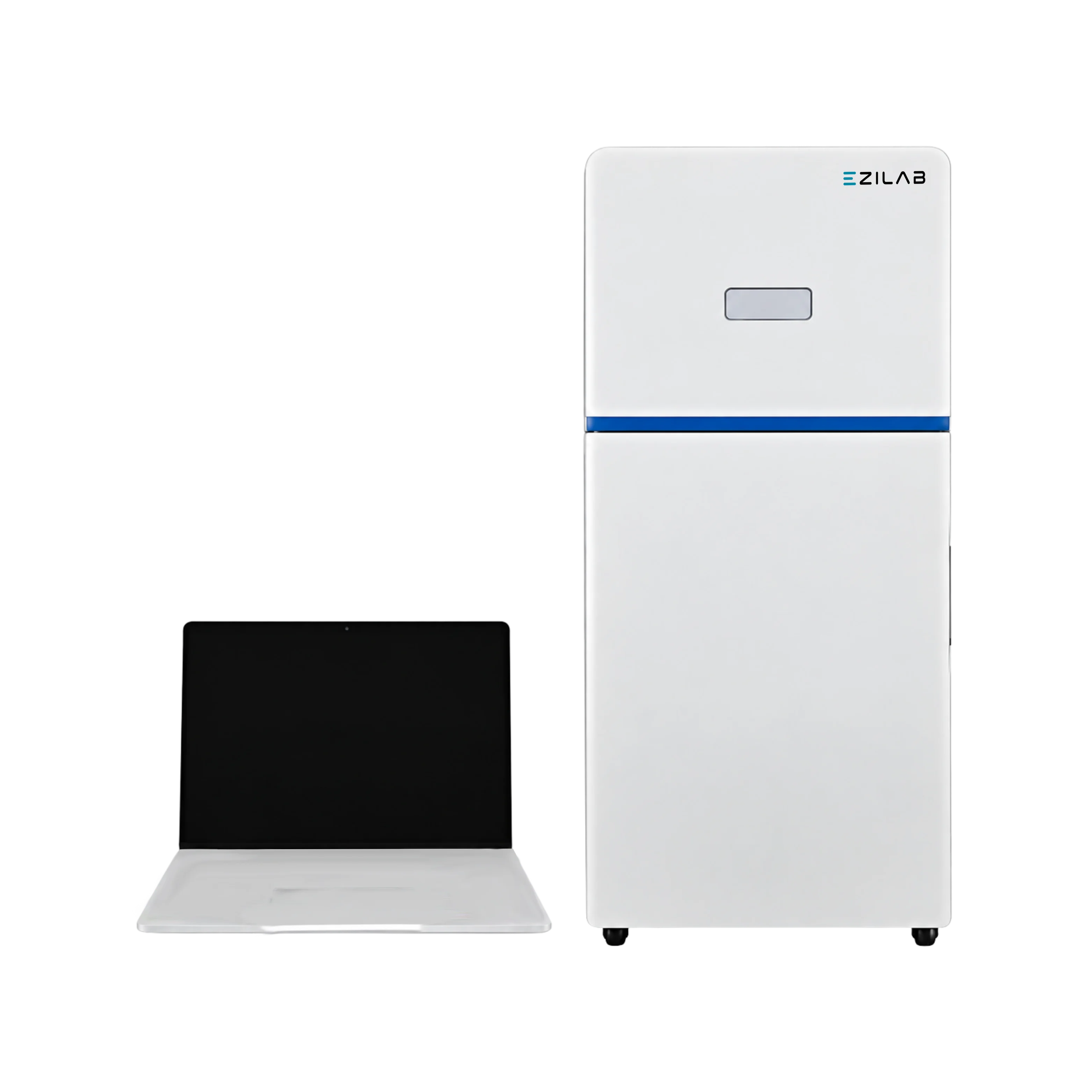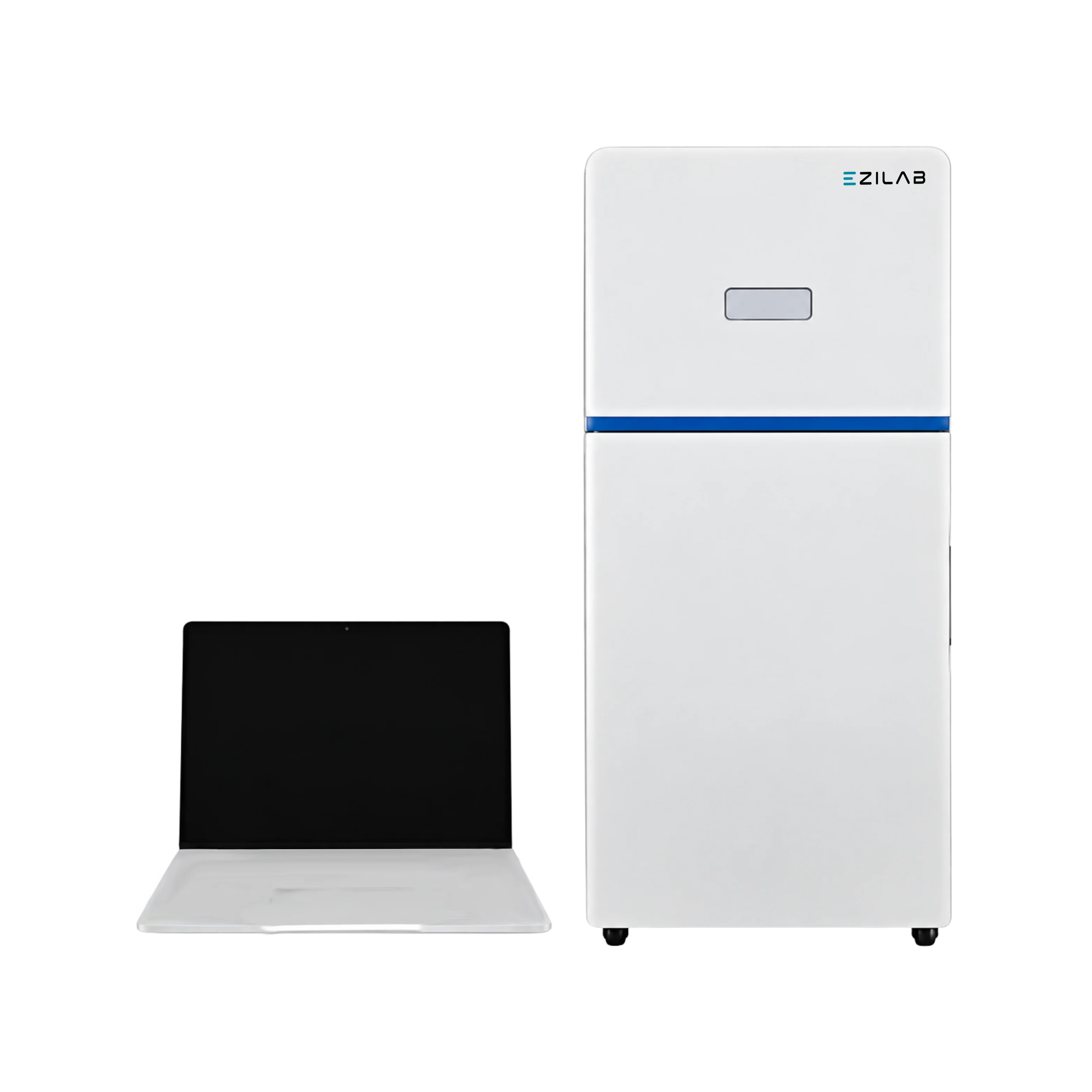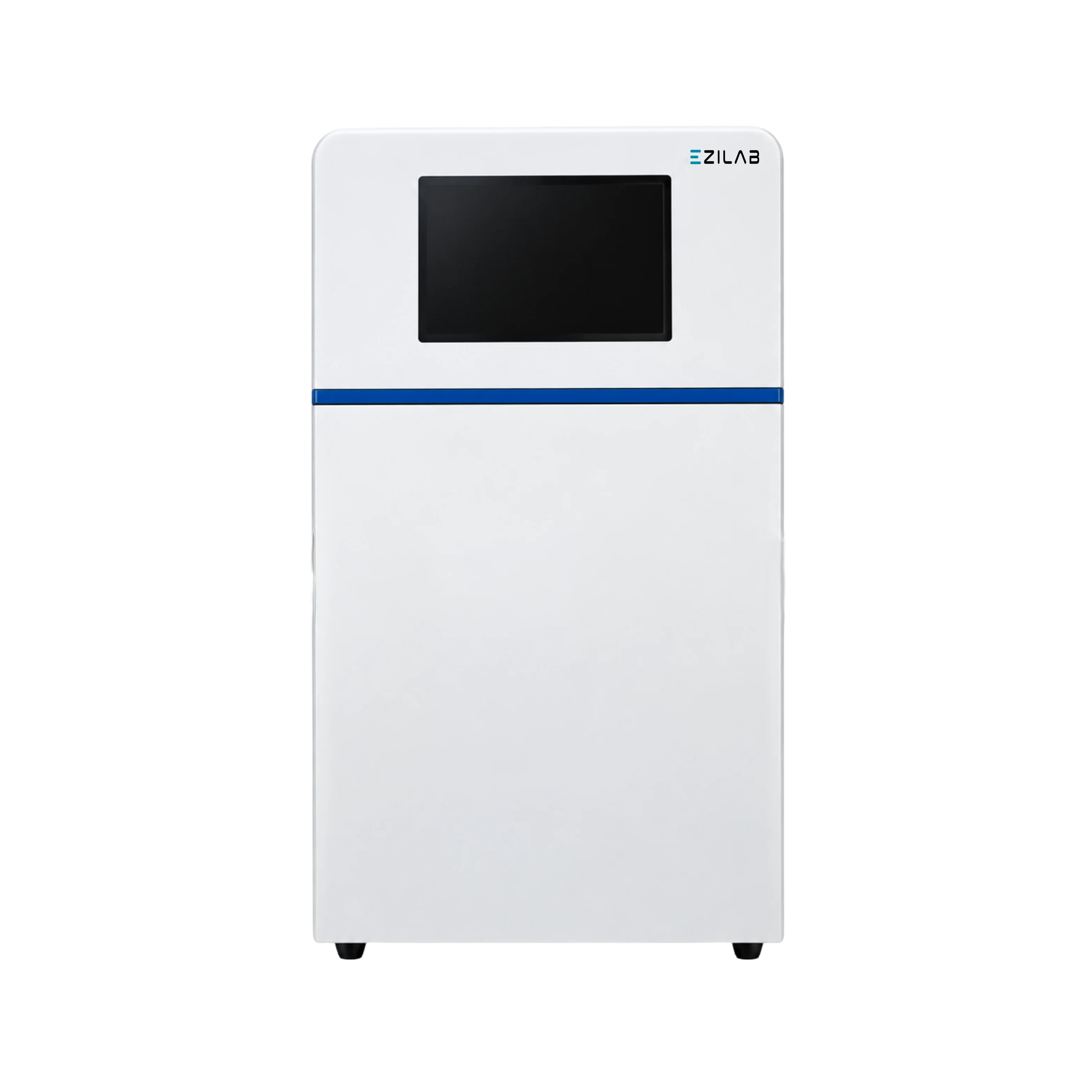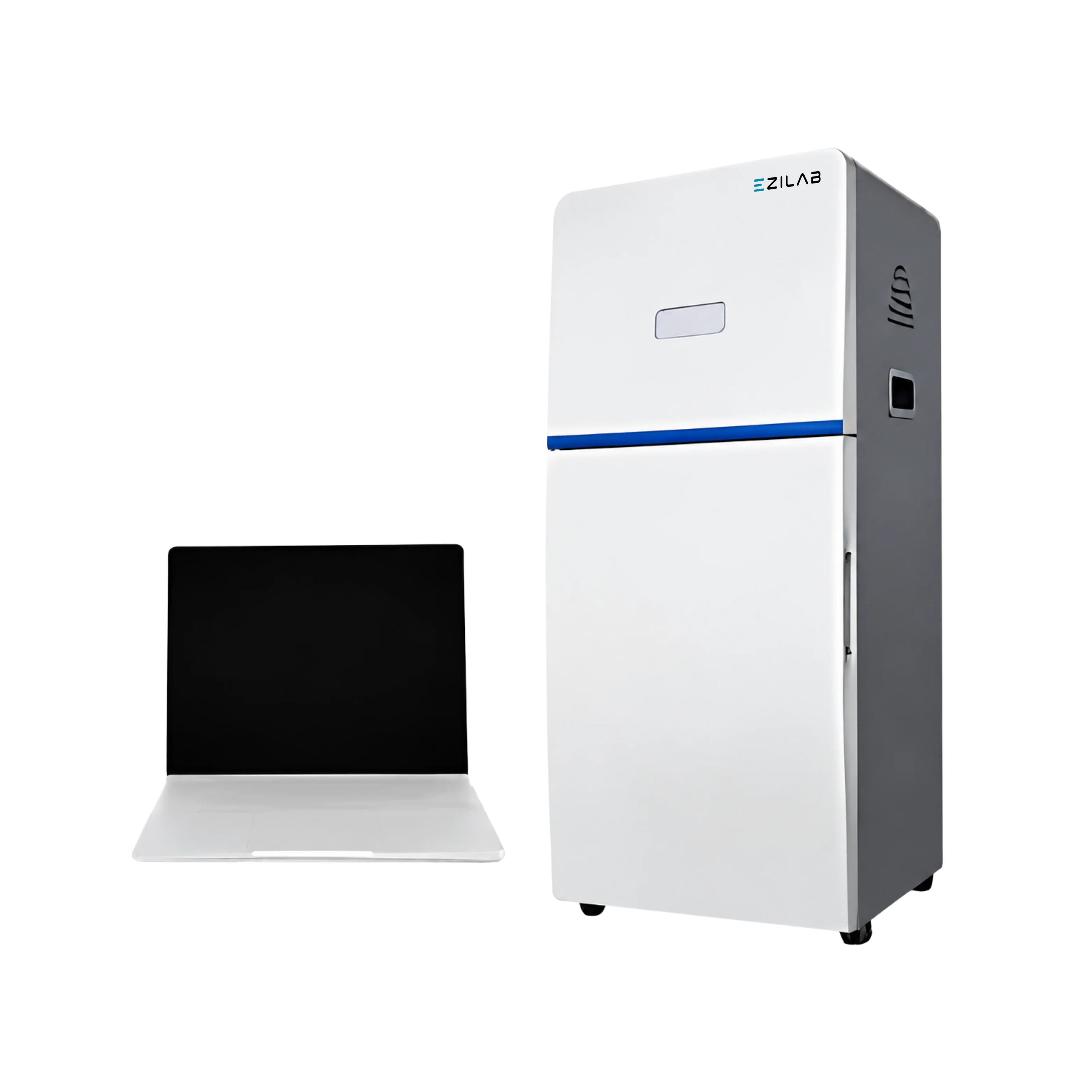
EziLab offers the Chemiluminescence Imaging System EZL-CS134, a gel imager and western blot imager built around a scientific CCD camera with 6.05 megapixels (2750 × 2200) and 16-bit, 65,536-level grayscale. The sensor cools to an absolute -25°C and reaches ≥75% quantum efficiency, so faint chemiluminescent signals show up with low background noise. A motorized F0.8 ultrafast lens gathers more light, pixel binning boosts sensitivity for weak bands, and automatic exposure sets capture time for each sample. An ultra-slim trans illuminator offers blue and white light for stained gels and colored samples. The 10.1-inch touchscreen runs capture and analysis, with automatic gel band recognition and optical density calculation for quantitative results. The benchtop cabinet measures 430 × 380 × 690 mm. As a chemiluminescence imager, it captures Western blots with ECL substrates, DNA and RNA gels, and protein gels in one dark cabinet, replacing film and darkroom work. Molecular biology labs document PCR products and nucleic acid gels, while proteomics and cell biology teams measure protein expression on blotting membranes. Biotech, pharmaceutical, and clinical research labs quantify target proteins, and universities use the system for teaching and research. See the Chemiluminescence Imaging Systems and Chemiluminescence Imaging System pages for full specifications.
| Lens | Motorized F0.8 Ultrafast Lens |
|---|---|
| Touch Screen | 10.1 Inch Touch Screen |
| Camera | Scientific CCD Camera |
| Resolution | 6.05 Megapixels, 2750×2200 |
| Sensor Size | 12.49mm ×9.99mm |
| Pixel Density | 16 bit ( 65536 Grayscale) |
| Pixel Size | 4.54×4.54µm |
| Quantum Efficiency | ≥ 75% |
| Absolute Cooling Temperature | -25°C |
| Product Dimension (W×D×H) | 430×380×690 mm |
| Outer Pack Dimension (W×D×H) | 550×490×760 mm |
| Gross Weight | 31 kg |
Ultra slim trans illuminator
Front and door are designed by reaction injection molding
Better user experience
Switchable operation mode
Blue and White light trans illuminator
Intelligent workflow
User interface allows total customization over imaging protocols
High precision automatic exposure
Marker image capture and composition with objective brands
Convenient image navigation and browsing
Automatic recognition of gel bands
Optimization of the visual effects by wiping background mode
Advanced pixel binning technology
Calculation of optical density for the quantitative analysis
This system is widely used for Gel inspection, Protein inspection, Blotting Membrane, Color imaging and DNA/RNA analysis.